
Bassett Collection of Stereoscopic Images of Human Anatomy
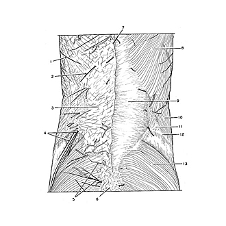
Dissection of thoracic and lumbosacral regions of back from a posterior approach
Superficial structures and external layer of muscles of lumbosacral region of back
Image #212-3
KEYWORDS: Lumbar region, Muscles and tendons, Peripheral nervous system, Sacral region, Thoracic region, Vasculature, Vertebral column.
Creative Commons
Stanford holds the copyright to the David L. Bassett anatomical images and has assigned Creative Commons license Attribution-Share Alike 4.0 International to all of the images.

For additional information regarding use and permissions, please contact the Medical History Center.

Dissection of thoracic and lumbosacral regions of back from a posterior approach
Superficial structures and external layer of muscles of lumbosacral region of back
The lower part of the specimen is shown in view 212-1 is shown in more detail in this close-up photograph.
- Lateral cutaneous branch, intercostal nerve VII (posterior branch)
- Lateral cutaneous branch of dorsal branch thoracic nerve XI
- Superficial fascia
- Superior cluneal nerves
- Middle cluneal nerves
- Coccyx (covered by ligaments)
- Middle cutaneous branch of dorsal branch thoracic nerve X
- Latissimus dorsi muscle
- Thoracolumbar fascia (overlying erector spinae muscle)
- External oblique muscle
- Lumbar triangle (Petit's triangle)
- Iliac crest
- Gluteus maximus muscle