Bassett Collection of Stereoscopic Images of Human Anatomy
Exploration of a brain cut in frontal section at the splenium of the corpus callosum
Anterior medullary velum, trochlear nerve, inferior and superior colliculi and pineal body
Image #21-5
KEYWORDS: Brain, Diencephalon, Medulla, Telencephalon, Vasculature.
Creative Commons
Stanford holds the copyright to the David L. Bassett anatomical images and has assigned Creative Commons license Attribution-Share Alike 4.0 International to all of the images.

For additional information regarding use and permissions, please contact the Medical History Center.
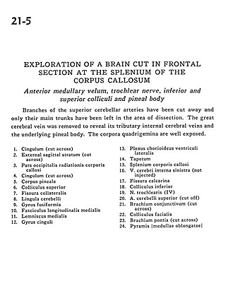
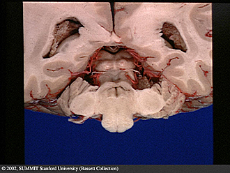
Exploration of a brain cut in frontal section at the splenium of the corpus callosum
Anterior medullary velum, trochlear nerve, inferior and superior colliculi and pineal body
Branches of the superior cerebellar arteries have been cut away and only their main trunks have been left in the area of dissection. The great cerebral vein was removed to reveal its tributary internal cerebral veins and the underlying pineal body. The corpora quadrigemina are well exposed.
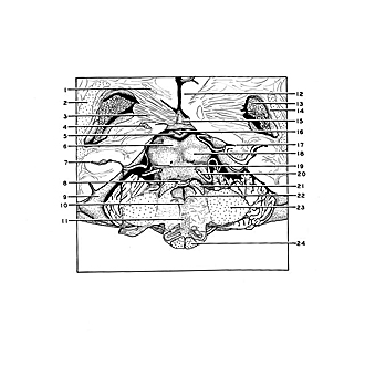
- Cingulum (cut across)
- External sagittal stratum (cut across)
- Occipital part of radiations of corpus callosum
- Cingulum (cut across)
- Pineal body
- Superior colliculus
- Collateral fissure
- Cerebellar lingula
- Fusiform gyrus
- Medial longitudinal fasciculus
- Medial lemniscus
- Cingulate gyrus
- Choroid plexus lateral ventricle
- Tapetum
- Corpus callosum (splenium)
- Internal cerebral vein left (not injected)
- Calcarine fissure
- Inferior colliculus
- Trochlear nerve (IV)
- Superior cerebellar artery (cut off)
- Brachium conjunctivum (superior cerebellar peduncle) (cut across)
- Facial colliculus
- Brachium pontis (middle cerebellar peduncle) (cut across)
- Pyramis of medulla oblongota