Bassett Collection of Stereoscopic Images of Human Anatomy
Exploration of a brain cut in frontal section at the splenium of the corpus callosum
Orientation view, cerebellum partly resected
Image #21-4
KEYWORDS: Brain, Cerebellum, Medulla, Vasculature, Ventricules.
Creative Commons
Stanford holds the copyright to the David L. Bassett anatomical images and has assigned Creative Commons license Attribution-Share Alike 4.0 International to all of the images.

For additional information regarding use and permissions, please contact the Medical History Center.

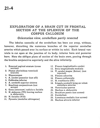
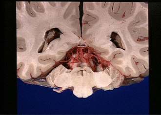
Exploration of a brain cut in frontal section at the splenium of the corpus callosum
Orientation view, cerebellum partly resected
The lobulus centralis of the cerebellum has been cut away, without, however, disturbing the numerous branches of the superior cerebellar arteries which passed over its surface or within its sulci. Each lateral ventricle is cut open at the junction of its body, inferior horn and posterior horn. Note the oblique plane of section of the brain stem, passing through the brachia conjunctiva superiorly and the olive inferiorly.
- External sagittal stratum (cross section)
- Choroid plexus lateral ventricle
- Hippocampus
- Posterior cerebral artery (cut off)
- Superior colliculus
- Superior cerebellar artery left
- Brachium conjunctivum (superior cerebellar peduncle) (cut across)
- Roots of facial nerve
- Abducens nerve (VI) leaving nucleus of abducens nerve
- Facial nerve (VII)
- Pyramis [medulla oblongata]
- Longitudinal fissure (cerebral)
- Medial longitudinal stria
- Great cerebral vein (of Galen) (not injected)
- Calcarine fissure
- Collateral fissure
- Trochlear nerve (IV)
- Cerebellar lingula lying on anterior medullary velum
- Fourth ventricle
- Nucleus of abducens nerve
- Brachium pontis (middle cerebellar peduncle) (cut across)
- Cerebellum
- Medial lemniscus (cut across)
- Inferior olivary nucleus