Bassett Collection of Stereoscopic Images of Human Anatomy
Radiography
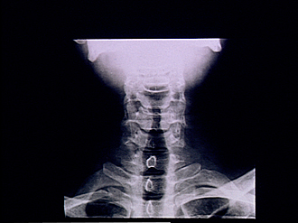
Radiograph of neck, anteroposterior view
Image #209-6
KEYWORDS: Bones joints cartilage, Cervical region, Vertebral column.
Creative Commons
Stanford holds the copyright to the David L. Bassett anatomical images and has assigned Creative Commons license Attribution-Share Alike 4.0 International to all of the images.

For additional information regarding use and permissions, please contact the Medical History Center.

Radiography
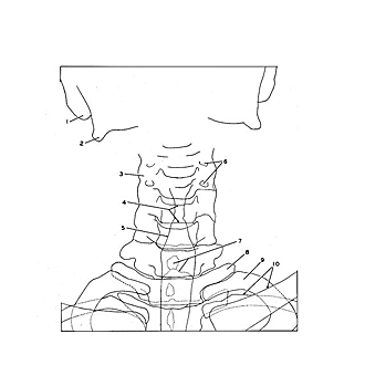
Radiograph of neck, anteroposterior view
The head has been extended. As a result the superimposed shadow of the base of the skull has obscured the upper three cervical vertebrae. Structural details of the transverse processes and articular processes of the cervical vertebrae are indistinct because of the overlapping of multiple shadows. The cavities of the larynx and trachea are outlined by their content of air. The conus elasticus of the larynx is indicated at 5. This film was obtained through the courtesy of Dr. Melvin J. Figley.
- Mastoid process
- Angle of mandible
- Area of overlapping shadows of articular processes and transverse processes
- Body of vertebra C. V (pointers indicate upper and lower borders of body)
- Trachea (pointer indicates wall of air-filled conus elasticus)
- Transverse foramen
- Upper pointer: Spinous process vertebra C. Vll Lower pointer: Lamina (arch of vertebra) C. VII
- Transverse process vertebra T. I
- Rib I
- Left pointer: Rib II Right pointer: Clavicle (in foreground)