Bassett Collection of Stereoscopic Images of Human Anatomy
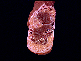
Cross sections of right foot
Section through sustentaculum tali and tarsal sinus
Image #205-3
KEYWORDS: Ankle, Bones joints cartilage, Foot and toes.
Creative Commons
Stanford holds the copyright to the David L. Bassett anatomical images and has assigned Creative Commons license Attribution-Share Alike 4.0 International to all of the images.

For additional information regarding use and permissions, please contact the Medical History Center.
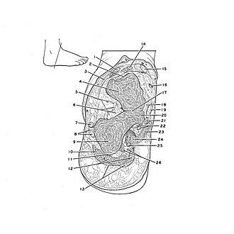
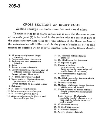
Cross sections of right foot
Section through sustentaculum tali and tarsal sinus
The plane of the cut is nearly vertical and is such that the anterior part of the ankle joint (2) is included in the section with the posterior part of the talocalcaneonavicular joint (17). The relation of the flexor tendons to the sustentaculum tali is illustrated. In the plane of section all of the long tendons are enclosed within synovial sheaths reinforced by fibrous sheaths.
- Extensor digitorum longus muscle (tendon)
- Talocrural articular space
- Inferior extensor retinaculum
- Talus
- Branch of lateral tarsal artery
- Upper pointer: Intemediate root of inferior extensor retinaculum Lower pointer: Tarsal sinus
- Peroneus brevis muscle (tendon)
- Upper pointer: Inferior peroneal retinaculum Lower pointer: Peroneus longus muscle
- Calcaneus
- Abductor digiti minimi muscle
- Long plantar ligament
- Flexor digitorum brevis muscle
- Plantar aponeurosis (lateral and intermedial portions)
- Extensor hallucis longus muscle (tendon)
- Tibialis anterior muscle (tendon)
- Greater saphenous vein
- Talocalcaneonavicular articular space (articulation between talus and middle surface of calcaneus)
- Tibiocalcaneal part of deltoid ligament
- Tibialis posterior muscle (tendon within synovial sheath)
- Sustentaculum tali
- Flexor digitorum longus muscle (tendon within synovial sheath)
- Upper pointer: Flexor hallucis longus muscle (tendon within synovial sheath) Lower pointer: Medial plantar nerve (medial plantar artery lies slightly inferior to nerve and is small)
- Abductor hallucis muscle
- Quadratus plantae muscle
- Lateral plantar nerve
- Lateral plantar artery and vein