
Bassett Collection of Stereoscopic Images of Human Anatomy
Cross sections of right foot
Section through proximal parts of metatarsal shafts, viewed from in front
Image #204-6
KEYWORDS: Bones joints cartilage, Foot and toes.
Creative Commons
Stanford holds the copyright to the David L. Bassett anatomical images and has assigned Creative Commons license Attribution-Share Alike 4.0 International to all of the images.

For additional information regarding use and permissions, please contact the Medical History Center.
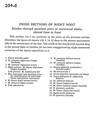
Cross sections of right foot
Section through proximal parts of metatarsal shafts, viewed from in front
This section, cut 2 cm. posterior to the plane of the previous section, illustrates the layers of the muscle (19, 4, 14, 5) deep to the plantar aponeurosis (20) in the central part of the foot. The width of the fascial cleft located deep to the second layer of muscles (4) has been exaggerated by slight downward retraction of the layers superficial to it.
- Fascia of dorsalis pedis
- Extensor digitorum longus muscle (tendons)
- Extensor digitorum brevis muscle
- Right pointer: Flexor digitorum longus muscle (tendon) Left pointer: 3rd lumbrical muscle
- Interosseous muscle (see previous view for identification of individual dorsal and plantar interossei)
- Flexor digiti minimi brevis muscle
- Abductor digiti minimi muscle
- Tela subcutanea
- Extensor hallucis brevis muscle (tendon)
- Extensor hallucis longus muscle (tendon)
- Deep plantar branch of dorsalis pedis artery
- Metatarsal bone
- Plantar arch (partially cut away)
- Oblique head of adductor hallucis muscle
- Abductor hallucis muscle
- Flexor hallucis brevis muscle
- Flexor hallucis longus muscle (tendon)
- Medial plantar nerve
- Flexor digitorum brevis muscle
- Plantar aponeurosis
- Lateral plantar nerve