
Bassett Collection of Stereoscopic Images of Human Anatomy
Joints of left ankle and foot
Interior of ankle joint, anterior view
Image #203-7
KEYWORDS: Ankle, Bones joints cartilage, Foot and toes.
Creative Commons
Stanford holds the copyright to the David L. Bassett anatomical images and has assigned Creative Commons license Attribution-Share Alike 4.0 International to all of the images.

For additional information regarding use and permissions, please contact the Medical History Center.
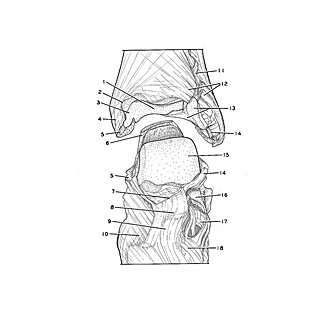
Joints of left ankle and foot
Interior of ankle joint, anterior view
The tibia and fibula have been detached from the tarsal bones. The foot has been placed in a position of partial extension.
- Inferior articular surface of tibia
- Talocrural articular capsule (cut edge)
- Malleolar articular surface
- Medial malleolus
- Deltoid ligament (divided)
- Tendo calcaneus
- Neck of talus (pointer on cut margin of joint capsule)
- Head of talus (covered by ligaments)
- Talonavicular ligament
- Navicular bone (covered by ligaments)
- Interosseous tibiofibular ligament
- Anterior tibiofibular ligament (divided)
- Upper pointer: Malleolar articular surface Lower pointer: Transverse tibiofibular ligament
- Anterior talofibular ligament (divided)
- Superior surface of trochlea of talus
- Tarsal sinus
- Calcaneus (pointer on superior tubercle)
- Dorsal cuboideonavicular ligament