Bassett Collection of Stereoscopic Images of Human Anatomy
Joints of left ankle and foot
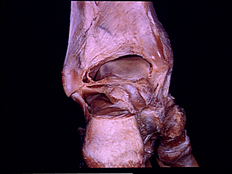
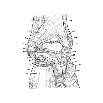
Interior of ankle joint and subtalar joint in relation to deep ligaments, posterior view
Image #203-6
KEYWORDS: Ankle, Bones joints cartilage, Foot and toes, Muscles and tendons.
Creative Commons
Stanford holds the copyright to the David L. Bassett anatomical images and has assigned Creative Commons license Attribution-Share Alike 4.0 International to all of the images.

For additional information regarding use and permissions, please contact the Medical History Center.

Joints of left ankle and foot
Interior of ankle joint and subtalar joint in relation to deep ligaments, posterior view
The joint capsules have been widely opened. All of the major ligaments of the ankle joint and subtalar joint have been kept intact for the purpose of this photograph. In the following views the bones have been separated by transection of the ligaments.
- Fibula
- Posterior tibiofibular ligament
- Upper pointer: Transverse tibiofibular ligament Lower pointer: Lateral malleolar surface trochlea of talus
- Talocrural articular capsule (cut edge of capsule traceable as thin ridge of tissue around margin of opened joint cavity)
- Lateral malleolus
- Posterior talofibular ligament (note massive size and depth of ligament)
- Calcaneofibular ligament
- Upper pointer: Posterior articular surface of calcaneus for talus Lower pointer: Calcaneus
- Upper pointer: Lateral tubercle of posterior talar process Lower pointer: Subtalar articular capsule (posterior talocalcaneal ligament)
- Upper pointer: Groove for flexor hallucis longus tendon Lower pointer: Medial tubercle of posterior talar process
- Tuberosity of calcaneus
- Tibia
- Superior surface of trochlea of talus
- Malleolar groove (for tibialis posterior muscle)
- Medial malleolus
- Posterior tibiotalar part of deltoid ligament
- Tibiocalcaneal part of deltoid ligament
- Medial talocalcaneal ligament (fibers run horizontally)
- Groove for flexor hallucis longus tendon
- Sustentaculum tali
- Tuberosity of navicular bone (tendon of tibialis posterior cut off at attachment)