
Bassett Collection of Stereoscopic Images of Human Anatomy
Joints of left ankle and foot
Interior of ankle and mid-tarsal joints in relation to ligaments, lateral view
Image #203-3
KEYWORDS: Ankle, Bones joints cartilage, Foot and toes, Muscles and tendons.
Creative Commons
Stanford holds the copyright to the David L. Bassett anatomical images and has assigned Creative Commons license Attribution-Share Alike 4.0 International to all of the images.

For additional information regarding use and permissions, please contact the Medical History Center.
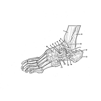

Joints of left ankle and foot
Interior of ankle and mid-tarsal joints in relation to ligaments, lateral view
The thin parts of the capsules of the ankles and mid-tarsal joints have been cut away to emphasize the position of the ligaments in relation to the bony and cartilaginous parts of these joints.
- Tibia
- Trochlea of talus (pointer on superior surface)
- Anterior talofibular ligament
- Cervical ligament
- Talonavicular ligament
- Calcaneonavicular ligament (part of bifurcate ligament)
- Calcaneocuboid ligament (part of bifurcate ligament)
- Dorsal cuneonavicular ligament
- Fibula
- Anterior tibiofibular ligament
- Lateral process of talus
- Left pointer: Lateral talocalcaneal ligament Right pointer: Calcaneofibular ligament
- Calcaneus
- Inferior peroneal retinaculum (opened)
- Left pointer: Inferior extensor retinaculum (intermediate root) Right pointer: Tarsal sinus (leading into deeply placed tarsal canal)
- Dorsal calcaneocuboid ligament
- Dorsal cuboideonavicular ligament