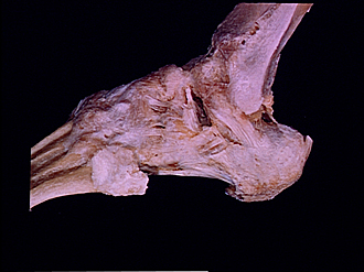
Bassett Collection of Stereoscopic Images of Human Anatomy
Joints of left ankle and foot
Ligaments of ankle and foot, lateral view
Image #203-2
KEYWORDS: Ankle, Bones joints cartilage, Foot and toes, Muscles and tendons.
Creative Commons
Stanford holds the copyright to the David L. Bassett anatomical images and has assigned Creative Commons license Attribution-Share Alike 4.0 International to all of the images.

For additional information regarding use and permissions, please contact the Medical History Center.

Joints of left ankle and foot
Ligaments of ankle and foot, lateral view
- Tibia
- Anterior tibiofibular ligament
- Talocrural articular capsule
- Anterior talofibular ligament
- Tarsal canal in depths of tarsal sinus (pointer on medial root of inferior extensor retinaculum)
- Head of talus
- Cervical ligament
- Talonavicular ligament
- Navicular bone (covered by ligaments)
- Bifurcate ligament (pointer on calcaneonavicular ligament; calcaneocuboid ligament not visible in dissection)
- Dorsal cuboideonavicular ligament
- Dorsal cuneonavicular ligament
- Fibula
- Lateral malleolus
- Subtalar articular capsule
- Upper pointer: Lateral talocalcaneal ligament Lower pointer: Calcaneofibular ligament
- Tendo calcaneus
- Calcaneus
- Lateral tubercle process of calcaneus bone
- Medial processs (plantar aponeurosis cut across)
- Inferior peroneal retinaculum (opened)
- Inferior extensor retinaculum (upper pointer, intermediate root; lower pointer, lateral root)
- Superior tubercle of calcaneus
- Dorsal calcaneocuboid ligament
- Long plantar ligament
- Cuboid bone (covered by ligaments)
- Peroneus brevis muscle (tendon cut off at insertion)
- Dorsal cuneocuboid ligament
- Dorsal metatarsal ligament