
Bassett Collection of Stereoscopic Images of Human Anatomy
Dissection of plantar aspect of left foot
Interosseous muscles and plantar arch, close-up plantar view
Image #202-3
KEYWORDS: Foot and toes, Muscles and tendons, Peripheral nervous system, Vasculature.
Creative Commons
Stanford holds the copyright to the David L. Bassett anatomical images and has assigned Creative Commons license Attribution-Share Alike 4.0 International to all of the images.

For additional information regarding use and permissions, please contact the Medical History Center.

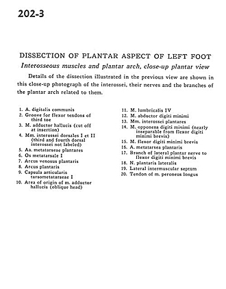
Dissection of plantar aspect of left foot
Interosseous muscles and plantar arch, close-up plantar view
Details of the dissection illustrated in the previous view are shown in this close-up photograph of the interossei, their nerves and the branches of the plantar arch related to them.
- Common digital artery
- Groove for flexor tendons of third toe
- Adductor hallucis muscle (cut off at insertion)
- 1st and 2nd dorsal interosseous muscles (3rd and 4th dorsal interossei not labeled)
- Metatarsal plantar arteries
- Metatarsal bone
- Plantar venous arch
- Plantar arch
- 1st tarsometatarsal articular capsule
- Area of origin of adductor hallucis muscle (oblique head)
- 4th lumbrical muscle
- Abductor digiti minimi muscle
- Plantar interosseous muscles
- Opponens digiti minimi muscle (nearly inseparable from flexor digiti minimi brevis)
- Flexor digiti minimi brevis muscle
- Plantar metatarsal artery
- Branch of lateral plantar nerve to flexor digiti minimi brevis
- Lateral plantar nerve
- Lateral intermuscular septum
- Tendon of peroneus longus muscle