Bassett Collection of Stereoscopic Images of Human Anatomy
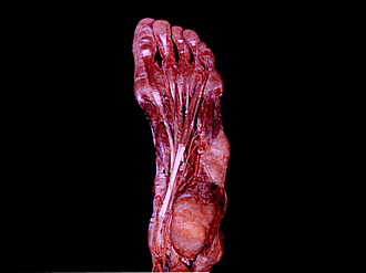
Dissection of plantar aspect of left foot
Long flexor tendons and lumbrical muscles
Image #201-6
KEYWORDS: Foot and toes, Muscles and tendons.
Creative Commons
Stanford holds the copyright to the David L. Bassett anatomical images and has assigned Creative Commons license Attribution-Share Alike 4.0 International to all of the images.

For additional information regarding use and permissions, please contact the Medical History Center.


Dissection of plantar aspect of left foot
Long flexor tendons and lumbrical muscles
The lateral tendons of the flexor digitorum longus have been slightly elevated.
- Common plantar digital artery
- Flexor hallucis longus muscle (tendon within opened digital sheath)
- Adductor hallucis muscle
- Flexor hallucis brevis muscle
- Deep branch lateral plantar nerve
- Area of origin of flexor digitorum brevis muscle and plantar aponeurosis
- Flexor digitorum longus muscle
- Flexor hallucis longus muscle (tendon within opened flexor retinaculum at ankle)
- Tuberosity of calcaneus
- Tibial nerve
- Lumbrical muscles
- Flexor digiti minimi brevis muscle
- Quadratus plantae muscle
- Lateral plantar artery
- Position of tendon of peroneus longus muscle
- Abductor digiti minimi muscle (cut off at origin)
- Accessory peroneus muscle
- Lateral malleolus (pointer on groove for peroneal tendons)