Bassett Collection of Stereoscopic Images of Human Anatomy
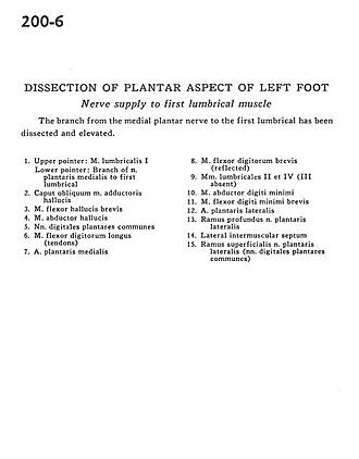
Dissection of plantar aspect of left foot
Nerve supply to first lumbrical muscle
Image #200-6
KEYWORDS: Foot and toes, Muscles and tendons, Peripheral nervous system.
Creative Commons
Stanford holds the copyright to the David L. Bassett anatomical images and has assigned Creative Commons license Attribution-Share Alike 4.0 International to all of the images.

For additional information regarding use and permissions, please contact the Medical History Center.
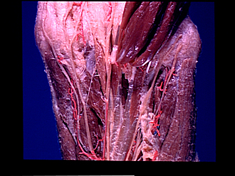
Dissection of plantar aspect of left foot
Nerve supply to first lumbrical muscle
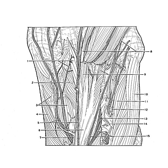
The branch from the medial plantar nerve to the first lumbrical has been dissected and elevated.
- Upper pointer: Lumbrical muscle I Lower pointer: Branch of medial plantar nerve to first lumbrical
- Oblique head of adductor hallucis muscle
- Flexor hallucis brevis muscle
- Abductor hallucis muscle
- Common plantar digital nerves
- Flexor digitorum longus muscle (tendons)
- Medial plantar artery
- Flexor digitorum brevis muscle (reflected)
- 2nd and 4th lumbrical muscles (3rd absent)
- Abductor digiti minimi muscle
- Flexor digiti minimi brevis muscle
- Lateral plantar artery
- Deep branch lateral plantar nerve
- Lateral intermuscular septum
- Superficial branch of lateral plantar nerve (common plantar digital nerves)