Bassett Collection of Stereoscopic Images of Human Anatomy
Exploration of those parts of the brain supplied by the posterior cerebral artery
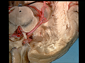
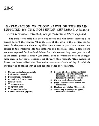
Stria terminalis reflected; temporothalamic fibers exposed
Image #20-6
KEYWORDS: Brain, Diencephalon, Telencephalon, Temporal lobe.
Creative Commons
Stanford holds the copyright to the David L. Bassett anatomical images and has assigned Creative Commons license Attribution-Share Alike 4.0 International to all of the images.

For additional information regarding use and permissions, please contact the Medical History Center.

Exploration of those parts of the brain supplied by the posterior cerebral artery
Stria terminalis reflected; temporothalamic fibers exposed
The stria terminalis has been cut across and the lower segment (12) turned toward the viewer. Thus the size of the stria in this region can be seen. In the previous view many fibers were seen to pass from the stratum zonale of the thalamus into the temporal and occipital lobes. These fibers are now exposed far into both lobes. In their course they pass just lateral to the lateral geniculate body (the lateral zone of Wernicke or area triangularis seen in horizontal sections cut through this region). This system of fibers has been called the "fasciculus temporothalamicus" by Arnold although it is apparent that it also reaches other cortical areas.
- Medial geniculate body
- Cerebral peduncle
- Interpeduncular fossa
- Basilar artery (cut across)
- Infundibulum
- Optic tract
- Optic nerve (II)
- Olfactory tract
- Lateral fissure (Sylvian)
- System of fibers extending from stratum zonale thalami into temporal and occipital lobes, the temporothalamic fasciculus
- Lateral geniculate body
- Stria terminalis (cut across and reflected)
- Amygdaloid nucleus (dissected)
- Medullary substance of Inferior temporal gyrus