Bassett Collection of Stereoscopic Images of Human Anatomy
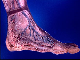
Dissection of medial aspect of left foot and ankle
Superficial nerves and blood vessels of foot, medial view
Image #198-5
KEYWORDS: Ankle, Foot and toes, Peripheral nervous system, Vasculature.
Creative Commons
Stanford holds the copyright to the David L. Bassett anatomical images and has assigned Creative Commons license Attribution-Share Alike 4.0 International to all of the images.

For additional information regarding use and permissions, please contact the Medical History Center.


Dissection of medial aspect of left foot and ankle
Superficial nerves and blood vessels of foot, medial view
The tela subcutanea has been dissected to reveal the superficial nerves, arteries and veins of the medial aspect of the left foot. A margin of skin and subcutaneous tissue has been preserved at the borders of the dissected area. Copyright holder
- Medial malleolus
- Flexor retinaculum
- Medial calcaneal branches of tibial nerve
- Calcaneal branch of the posterior tibial artery
- Calcaneus
- Tela subcutanea
- Abductor hallucis muscle (covered by fascia)
- Plantar aponeurosis
- Greater saphenous vein
- Saphenous nerve
- Dorsal medial cutaneous nerve (note branch crossing superficial to saphenous nerve to reach plantar aspect of foot)
- Fascia overlying metatarsophalangeal joint