Bassett Collection of Stereoscopic Images of Human Anatomy
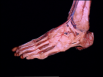
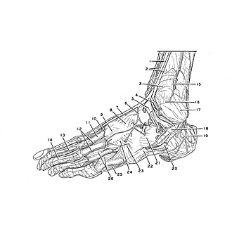
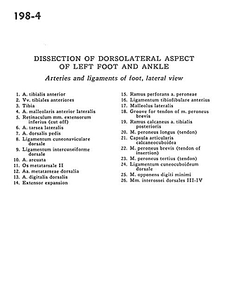
Dissection of dorsolateral aspect of left foot and ankle
Arteries and ligaments of foot, lateral view
Image #198-4
KEYWORDS: Ankle, Foot and toes, Muscles and tendons, Vasculature.
Creative Commons
Stanford holds the copyright to the David L. Bassett anatomical images and has assigned Creative Commons license Attribution-Share Alike 4.0 International to all of the images.

For additional information regarding use and permissions, please contact the Medical History Center.
Dissection of dorsolateral aspect of left foot and ankle
Arteries and ligaments of foot, lateral view
- Anterior tibial artery
- Anterior tibial veins
- Tibia
- Anterior lateral malleolar artery
- Inferior extensor retinaculum (cut off)
- Lateral tarsal artery
- Dorsalis pedis artery
- Dorsal cuneonavicular ligament
- Dorsal intercuneiform ligament
- Arcuate artery
- Metatarsal bone II
- Dorsal metatarsal arteries
- Dorsal digital artery
- Extensor expansion
- Perforating branch of peroneal artery
- Anterior tibiofibular ligament
- Lateral malleolus
- Groove for tendon of peroneus brevis muscle
- Calcaneal branch of the posterior tibial artery
- Peroneus longus muscle (tendon)
- Calcaneocuboid articular capsule
- Peroneus brevis muscle (tendon of insertion)
- Peroneus tertius muscle (tendon)
- Dorsal cuneocuboid ligament
- Opponens digiti minimi muscle
- 3rd-4th dorsal interosseous muscles