Bassett Collection of Stereoscopic Images of Human Anatomy
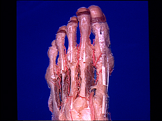
Dissection of dorsolateral aspect of left foot and ankle
Dorsal metatarsal arteries; dorsal interosseous muscles, viewed from above
Image #198-3
KEYWORDS: Ankle, Bones joints cartilage, Foot and toes, Muscles and tendons, Vasculature.
Creative Commons
Stanford holds the copyright to the David L. Bassett anatomical images and has assigned Creative Commons license Attribution-Share Alike 4.0 International to all of the images.

For additional information regarding use and permissions, please contact the Medical History Center.

Dissection of dorsolateral aspect of left foot and ankle
Dorsal metatarsal arteries; dorsal interosseous muscles, viewed from above
The dorsal interossei have been separated slightly from their bony origins to expose the metatarsal arteries. In this foot these vessels spring largely from the perforating arteries rather than from the arcuate artery (12) which is extremely small.
- Extensor expansion
- Plantar digital artery
- Dorsal digital artery
- Tendon of extensor digitorum longus muscle
- Tendon of extensor digitorum brevis muscle
- 1st-4th dorsal interosseous muscles
- Dorsal metatarsal arteries
- 5th metatarsal bone
- Fascial expansion covering metatarsophalangeal joint
- Extensor hallucis longus muscle (accessory tendon also present)
- Perforating branch of plantar metatarsal artery
- Upper pointer: Deep plantar branch of dorsalis pedis artery Lower pointer: Arcuate artery
- Dorsalis pedis artery