
Bassett Collection of Stereoscopic Images of Human Anatomy
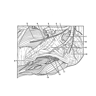
Dissection of dorsolateral aspect of left foot and ankle
Nerve supply to abductor digiti minimi muscle, close-up lateral view
Image #197-6
KEYWORDS: Ankle, Foot and toes, Muscles and tendons.
Creative Commons
Stanford holds the copyright to the David L. Bassett anatomical images and has assigned Creative Commons license Attribution-Share Alike 4.0 International to all of the images.

For additional information regarding use and permissions, please contact the Medical History Center.
Dissection of dorsolateral aspect of left foot and ankle
Nerve supply to abductor digiti minimi muscle, close-up lateral view
The abductor digiti minimi has been reflected downward to expose the branch of the lateral plantar nerve (12) as it passes within the substance of the muscle.
- Dorsal lateral cutaneous nerve
- Greater saphenous vein
- Peroneus longus muscle (tendon covered by inferior peroneal retinaculum)
- Peroneus brevis muscle (tendon covered by inferior peroneal retinaculum)
- Extensor digitorum brevis muscle
- Tuberosity of 5th metatarsal bone
- Accessory peroneus muscle
- Calcaneal branch of peroneal artery
- Inferior peroneal retinaculum
- Calcaneus (covered by periosteum)
- Branch of lateral plantar artery
- Branch of lateral plantar nerve (to abductor digiti minimi muscle)
- Abductor digiti minimi muscle (reflected downward)