
Bassett Collection of Stereoscopic Images of Human Anatomy
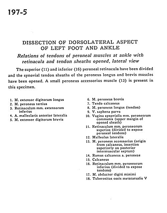
Dissection of dorsolateral aspect of left foot and ankle
Relations of tendons of peroneal muscles at ankle with retinacula and tendon sheaths opened, lateral view
Image #197-5
KEYWORDS: Ankle, Fascia, Foot and toes, Muscles and tendons.
Creative Commons
Stanford holds the copyright to the David L. Bassett anatomical images and has assigned Creative Commons license Attribution-Share Alike 4.0 International to all of the images.

For additional information regarding use and permissions, please contact the Medical History Center.

Dissection of dorsolateral aspect of left foot and ankle
Relations of tendons of peroneal muscles at ankle with retinacula and tendon sheaths opened, lateral view
The superior (11) and inferior (16) peroneal retinacula have been divided and synovial tendon sheaths of the peroneus longus and brevis muscles have been opened. A small peroneus accessorius muscle (13) is present in this specimen.
- Extensor digitorum longus muscle
- Peroneus tertius muscle
- Inferior extensor retinaculum
- Anterior lateral malleolar artery
- Extensor digitorum brevis muscle
- Peroneus brevis muscle
- Tendo calcaneus
- Peroneus longus muscle (tendon)
- Lesser saphenous vein
- Synovial sheath of peroneal muscles (upper margin of opened sheath)
- Superior peroneal retinaculum (divided to expose peroneal tendons)
- Lateral malleolus
- Accessory peroneus muscle (origin from calcaneus, insertion superiorly on posterior intermuscular septum)
- Calcaneal branch of peroneal artery
- Calcaneus
- Inferior peroneal retinaculum (divided to expose tendons)
- Abductor digiti minimi muscle
- Tuberosity of 5th metatarsal bone