Bassett Collection of Stereoscopic Images of Human Anatomy
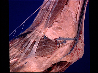
Dissection of dorsolateral aspect of left foot and ankle
Extensor retinacula and tendon sheath of extensor digitorum longus and peroneus tertius opened, anterolateral view of foot
Image #197-4
KEYWORDS: Ankle, Fascia, Foot and toes, Muscles and tendons.
Creative Commons
Stanford holds the copyright to the David L. Bassett anatomical images and has assigned Creative Commons license Attribution-Share Alike 4.0 International to all of the images.

For additional information regarding use and permissions, please contact the Medical History Center.


Dissection of dorsolateral aspect of left foot and ankle
Extensor retinacula and tendon sheath of extensor digitorum longus and peroneus tertius opened, anterolateral view of foot
The superior and inferior extensor retinacula (1,6) have been cut away to reveal the underlying tendons.
- Superior extensor retinaculum (partially removed)
- Peroneus tertius muscle
- Extensor digitorum longus muscle (tendons exposed deep to retinaculum)
- Peroneus tertius muscle (tendon of insertion exposed deep to retinaculum)
- Tibialis anterior muscle (tendon covered by retinaculum)
- Inferior extensor retinaculum (divided and reflected)
- Tendinous sheath (opened above level of upper pointer)
- Extensor hallucis brevis muscle
- Fascia of dorsalis pedis
- Lesser saphenous vein
- Superior peroneal retinaculum
- Anterior lateral malleolar artery
- Dorsal lateral cutaneous nerve
- Inferior peroneal retinaculum
- Peroneus brevis muscle (tendon of insertion)
- Extensor digitorum brevis muscle