Bassett Collection of Stereoscopic Images of Human Anatomy
Dissection of dorsolateral aspect of left foot and ankle
Synovial sheath of extensor tendons
Image #197-3
KEYWORDS: Ankle, Fascia, Foot and toes, Muscles and tendons.
Creative Commons
Stanford holds the copyright to the David L. Bassett anatomical images and has assigned Creative Commons license Attribution-Share Alike 4.0 International to all of the images.

For additional information regarding use and permissions, please contact the Medical History Center.
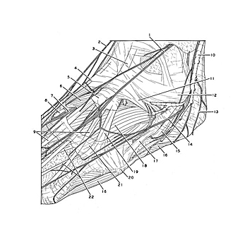
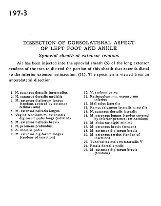
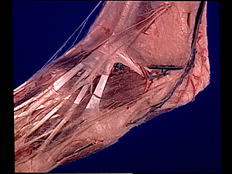
Dissection of dorsolateral aspect of left foot and ankle
Synovial sheath of extensor tendons
Air has been injected into the synovial sheath (5) of the long extensor tendons of the toes to distend the portion of this sheath that extends distal to the inferior extensor retinaculum (11). The specimen is viewed from an anterolateral direction.
- Dorsal intermediate cutaneous nerve
- Dorsal medial cutaneous nerve
- Extensor digitorum longus muscle (tendons covered by extensor retinaculum)
- Extensor hallucis longus muscle
- Tendinous sheath of extensor digitorum pedis longus muscle (inflated)
- Extensor hallucis brevis muscle
- Deep peroneal nerve
- Dorsalis pedis artery
- Extensor digitorum longus muscle (tendons of insertion)
- Lesser saphenous vein
- Inferior extensor retinaculum
- Lateral malleolus
- Calcaneal branch of lateral sural nerve
- Dorsal lateral cutaneous nerve
- Peroneus longus muscle (tendon covered by inferior peroneal retinaculum)
- Abductor digiti minimi muscle
- Peroneus brevis muscle (tendon)
- Extensor digitorum brevis muscle
- Peroneus tertius muscle (tendon of insertion)
- Tuberosity of 5th metatarsal bone
- Fascia of dorsalis pedis
- Extensor digitorum brevis muscle (tendons)