Bassett Collection of Stereoscopic Images of Human Anatomy
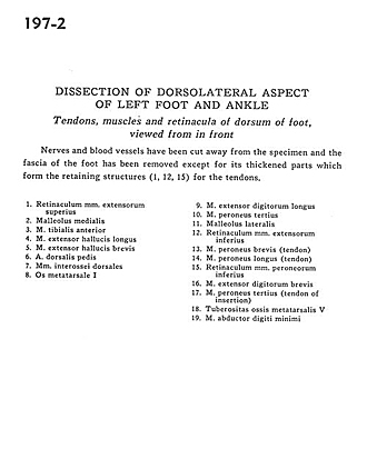
Dissection of dorsolateral aspect of left foot and ankle
Tendons, muscles and retinacula of dorsum of foot, viewed from in front
Image #197-2
KEYWORDS: Ankle, Foot and toes, Muscles and tendons.
Creative Commons
Stanford holds the copyright to the David L. Bassett anatomical images and has assigned Creative Commons license Attribution-Share Alike 4.0 International to all of the images.

For additional information regarding use and permissions, please contact the Medical History Center.
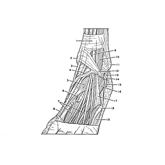
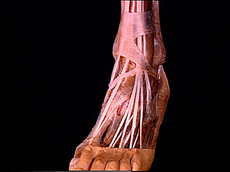
Dissection of dorsolateral aspect of left foot and ankle
Tendons, muscles and retinacula of dorsum of foot, viewed from in front
Nerves and blood vessels have been cut away from the specimen and the fascia of the foot has been removed except for its thickened parts which form the retaining structures (1,12,15) for the tendons.
- Superior extensor retinaculum
- Medial malleolus
- Tibialis anterior muscle
- Extensor hallucis longus muscle
- Extensor hallucis brevis muscle
- Dorsalis pedis artery
- Dorsal interosseous muscles
- metatarsal bone
- Extensor digitorum longus muscle
- Peroneus tertius muscle
- Lateral malleolus
- Inferior extensor retinaculum
- Peroneus brevis muscle (tendon)
- Peroneus longus muscle (tendon)
- Inferior peroneal retinaculum
- Extensor digitorum brevis muscle
- Peroneus tertius muscle (tendon of insertion)
- Tuberosity of 5th metatarsal bone
- Abductor digiti minimi muscle