Bassett Collection of Stereoscopic Images of Human Anatomy
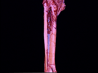
Dissection of posterior aspect of left leg
Interosseous membrane; area of origin of tibialis posterior muscle
Image #196-5
KEYWORDS: Leg, Muscles and tendons.
Creative Commons
Stanford holds the copyright to the David L. Bassett anatomical images and has assigned Creative Commons license Attribution-Share Alike 4.0 International to all of the images.

For additional information regarding use and permissions, please contact the Medical History Center.


Dissection of posterior aspect of left leg
Interosseous membrane; area of origin of tibialis posterior muscle
The tibialis posterior has been removed by dividing its fibres close to their origins (2) to demonstrate the interosseous membrane in relation to the posterior aspects of the tibia and fibula.
- Popliteal artery (at point of bifurcation into tibial vessels)
- Tibialis posterior muscle (area of origin)
- Fibula
- Interosseous membrane of leg
- Peroneal artery
- Popliteus muscle (area of insertion)
- Location of nutrient artery entering tibia
- Muscular and periosteal branch of tibialis posterior
- Tibia
- Communicating branch of peroneal artery