Bassett Collection of Stereoscopic Images of Human Anatomy
Dissection of posterior aspect of left leg
Nerves and blood vessels to tibialis posterior
Image #196-4
KEYWORDS: Leg, Muscles and tendons, Peripheral nervous system, Vasculature.
Creative Commons
Stanford holds the copyright to the David L. Bassett anatomical images and has assigned Creative Commons license Attribution-Share Alike 4.0 International to all of the images.

For additional information regarding use and permissions, please contact the Medical History Center.
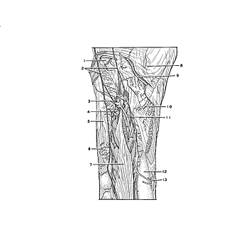
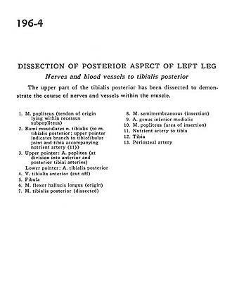
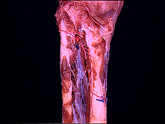
Dissection of posterior aspect of left leg
Nerves and blood vessels to tibialis posterior
The upper part of the tibialis posterior has been dissected to demonstrate the course of nerves and vessels within the muscle.
- Popliteus muscle (tendon of origin lying within subpopliteal recess)
- Muscular branch tibial nerve (to tibialis posterior muscle; upper pointer indicates branch to tibiofibular joint and tibia accompanying nutrient artery (II))
- Upper pointer: Popliteal artery (at division into anterior and posterior tibial arteries) Lower pointer: Posterior tibial artery
- Anterior tibial vein (cut off)
- Fibula
- Flexor hallucis longus muscle (origin)
- Tibialis posterior muscle (dissected)
- Semimembranosus muscle (insertion)
- Medial inferior genicular artery
- Popliteus muscle (area of insertion)
- Nutrient artery to tibia
- Tibia
- Periosteal artery