Bassett Collection of Stereoscopic Images of Human Anatomy
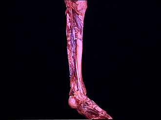
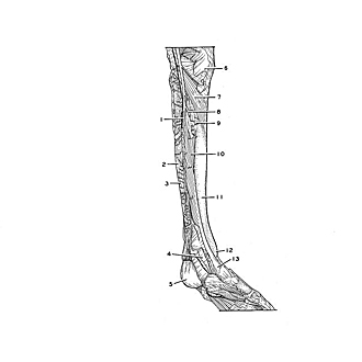
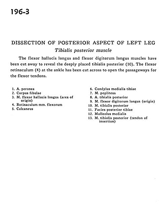
Dissection of posterior aspect of left leg
Tibialis posterior muscle
Image #196-3
KEYWORDS: Ankle, Leg, Muscles and tendons.
Creative Commons
Stanford holds the copyright to the David L. Bassett anatomical images and has assigned Creative Commons license Attribution-Share Alike 4.0 International to all of the images.

For additional information regarding use and permissions, please contact the Medical History Center.
Dissection of posterior aspect of left leg
Tibialis posterior muscle
The flexor hallucis longus and flexor digitorum longus muscles have been cut away to reveal the deeply placed tibialis posterior (10). The flexor retinaculum (4) at the ankle has been cut across to open the passageways for the flexor tendons.
- Peroneal artery
- Body of fibula
- Flexor hallucis longus muscle (area of origin)
- Flexor retinaculum
- Calcaneus
- Medial condyle of tibia
- Popliteus muscle
- Posterior tibial artery
- Flexor digitorum longus muscle (origin)
- Tibialis posterior muscle
- Posterior surface of tibia
- Medial malleolus
- Tibialis posterior muscle (tendon of insertion)