Bassett Collection of Stereoscopic Images of Human Anatomy
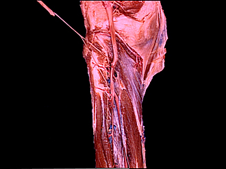
Dissection of posterior aspect of left leg
Popliteus muscle, close-up view
Image #196-1
KEYWORDS: Leg, Muscles and tendons.
Creative Commons
Stanford holds the copyright to the David L. Bassett anatomical images and has assigned Creative Commons license Attribution-Share Alike 4.0 International to all of the images.

For additional information regarding use and permissions, please contact the Medical History Center.


Dissection of posterior aspect of left leg
Popliteus muscle, close-up view
The heavy fascia that covered the popliteus has been cut away.
- Muscular branch of tibial nerve (stretched)
- Popliteus muscle (tendon of origin emerging through capsule of knee joint)
- Subpopliteal recess
- Muscular branch of tibial nerve (to tibialis posterior muscle)
- Muscular branch of tibial nerve (to popliteus muscle)
- Anterior tibial artery
- Posterior tibial artery
- Soleus muscle (origin)
- Peroneal artery
- Flexor hallucis longus muscle
- Tibialis posterior muscle
- Lateral head of gastrocnemius muscle
- Lateral inferior genicular artery
- Popliteal artery
- Oblique popliteal ligament
- Semimembranosus muscle (insertion)
- Medial inferior genicular artery
- Popliteus muscle
- Flexor digitorum longus muscle