Bassett Collection of Stereoscopic Images of Human Anatomy
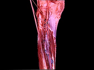
Dissection of posterior aspect of left leg
Nerves to deep flexor muscles, close-up view of upper area of leg
Image #195-7
KEYWORDS: Leg, Muscles and tendons, Peripheral nervous system.
Creative Commons
Stanford holds the copyright to the David L. Bassett anatomical images and has assigned Creative Commons license Attribution-Share Alike 4.0 International to all of the images.

For additional information regarding use and permissions, please contact the Medical History Center.
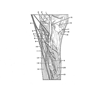

Dissection of posterior aspect of left leg
Nerves to deep flexor muscles, close-up view of upper area of leg
The tibial nerve has been elevated and put under traction to stretch the various muscular branches that have been exposed within the dissected area.
- Popliteal artery
- Oblique popliteal ligament
- Lateral inferior genicular artery
- Tibial nerve (elevated)
- Muscular branch Tibial nerve (to popliteus muscle and tibialis posterior muscle)
- Muscular branch of tibial nerve (to flexor digitorum longus muscle)
- Body of fibula
- Anterior tibial artery (passing into anterior compartment of leg)
- Posterior tibial artery
- Soleus muscle (origin)
- Muscular branch of tibial nerve (to flexor hallucis longus muscle)
- Peroneal artery
- Tibialis posterior muscle
- Flexor hallucis longus muscle
- Medial inferior genicular artery
- Semimembranosus muscle (insertion)
- Gracilis muscle (insertion)
- Sartorius muscle (insertion)
- Semitendinosus muscle (insertion)
- Flexor digitorum longus muscle
- Posterior tibial veins