
Bassett Collection of Stereoscopic Images of Human Anatomy
Dissection of posterior aspect of left leg
Flexor digitorum longus and flexor hallucis longus muscles; posterior tibial vessels; nerve supply to deep part of soleus muscle
Image #195-5
KEYWORDS: Leg, Muscles and tendons, Peripheral nervous system, Vasculature.
Creative Commons
Stanford holds the copyright to the David L. Bassett anatomical images and has assigned Creative Commons license Attribution-Share Alike 4.0 International to all of the images.

For additional information regarding use and permissions, please contact the Medical History Center.
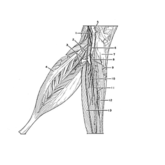

Dissection of posterior aspect of left leg
Flexor digitorum longus and flexor hallucis longus muscles; posterior tibial vessels; nerve supply to deep part of soleus muscle
The muscles and blood vessels within the deep compartment have been separated and more fully exposed than in the previous view. In addition, the small penniform component of the deep aspect of the soleus has been dissected to reveal the nerves and blood vessels that supply this portion of the muscle.
- Tibial nerve
- Muscular branch tibial nerve (to deep part of soleus)
- Sural arteries
- Soleus muscle
- Popliteal vein
- Muscular branch of tibial nerve (to flexor hallucis longus muscle)
- Popliteus muscle
- Posterior tibial artery
- Soleus muscle (origin from soleal line)
- Peroneal artery (accompanied by peroneal veins)
- Posterior tibial veins
- Flexor digitorum longus muscle
- Flexor hallucis longus muscle