
Bassett Collection of Stereoscopic Images of Human Anatomy
Dissection of posterior aspect of left leg
Plantaris muscle, close-up view of muscle belly and nerves supply
Image #195-3
KEYWORDS: Leg, Muscles and tendons, Peripheral nervous system.
Creative Commons
Stanford holds the copyright to the David L. Bassett anatomical images and has assigned Creative Commons license Attribution-Share Alike 4.0 International to all of the images.

For additional information regarding use and permissions, please contact the Medical History Center.
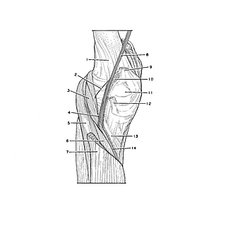
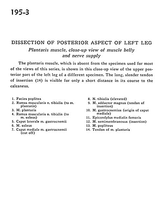
Dissection of posterior aspect of left leg
Plantaris muscle, close-up view of muscle belly and nerves supply
The plantaris muscle, which is absent from the specimen used for most of the views in this series, is shown in this close-up view of the upper posterior part of the left leg of a different specimen. The long, slender tendon of insertion (14) is visible for only a short distance in its course to the calcaneus.
- Popliteal surface
- Muscular branch of tibial nerve (to plantaris muscle)
- Plantaris muscle
- Muscular branch of tibial nerve (to soleus muscle)
- Lateral head of gastrocnemius muscle
- Soleus muscle
- Medial head of gastrocnemius muscle (cut off)
- Tibial nerve (elevated)
- Adductor magnus muscle (tendon of insertion)
- Gastrocnemius muscle (origin of medial head)
- Medial epicondyle of femur
- Semimembranosus muscle (insertion)
- Popliteus muscle
- Tendon of plantaris muscle