Bassett Collection of Stereoscopic Images of Human Anatomy
Dissection of posterior aspect of left leg
Bursa of semimembranosus muscle, close-up posterior view
Image #194-6
KEYWORDS: Leg, Muscles and tendons.
Creative Commons
Stanford holds the copyright to the David L. Bassett anatomical images and has assigned Creative Commons license Attribution-Share Alike 4.0 International to all of the images.

For additional information regarding use and permissions, please contact the Medical History Center.
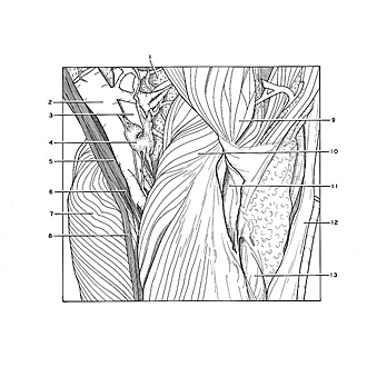
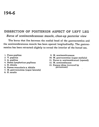
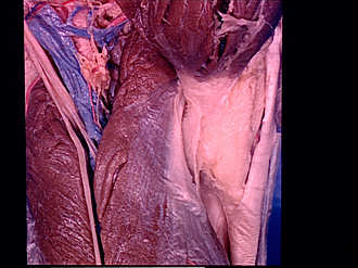
Dissection of posterior aspect of left leg
Bursa of semimembranosus muscle, close-up posterior view
The bursa that lies between the medial head of the gastrocnemius and semimembranosus muscle has been opened longitudinally. The gastrocnemius has been retracted slightly to reveal the interior of the bursal sac.
- Popliteal fossa
- Popliteal vein
- Popliteal artery
- Popliteal lymph node
- Tibial nerve
- Muscular branch of tibial nerve
- Gastrocnemius muscle (lateral head)
- Sural nerve
- Semimembranosus muscle
- Gastrocnemius muscle (medial head)
- Bursa of semimembranosus muscle (opened)
- Semitendinosus muscle
- Body of tibia (covered by periosteum)