Bassett Collection of Stereoscopic Images of Human Anatomy
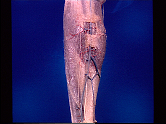
Dissection of posterior aspect of left leg
Superficial nerves and blood vessels of leg, posterior view
Image #194-3
KEYWORDS: Leg, Peripheral nervous system, Vasculature.
Creative Commons
Stanford holds the copyright to the David L. Bassett anatomical images and has assigned Creative Commons license Attribution-Share Alike 4.0 International to all of the images.

For additional information regarding use and permissions, please contact the Medical History Center.
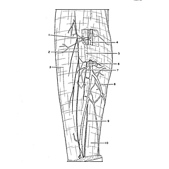
Dissection of posterior aspect of left leg
Superficial nerves and blood vessels of leg, posterior view
- Perforating vein (connecting with veins in gastrocnemius muscle)
- Lateral cutaneous sural nerve
- Crural fascia
- Lesser saphenous vein
- Tela subcutanea
- Cutaneous branch of posterior tibial artery
- Branch of medial sural cutaneous nerve
- Communication between large and small saphenous veins
- Sural nerve
- Tendo calcaneus (covered by fascia)