Bassett Collection of Stereoscopic Images of Human Anatomy
Dissection of medial aspect of left leg
Superficial nerves and blood vessels of medial side of leg
Image #194-1
KEYWORDS: Leg, Peripheral nervous system, Vasculature.
Creative Commons
Stanford holds the copyright to the David L. Bassett anatomical images and has assigned Creative Commons license Attribution-Share Alike 4.0 International to all of the images.

For additional information regarding use and permissions, please contact the Medical History Center.

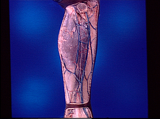
Dissection of medial aspect of left leg
Superficial nerves and blood vessels of medial side of leg
The tela subcutanea has been dissected. In the upper part of the field a small area of skin has been reflected upward to expose a cutaneous artery ramifying beneath the dermis.
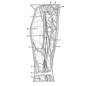
- Saphenous nerve
- Crural fascia (overlying triceps surae muscle)
- Accessory saphenous vein
- Communicating vein between greater and lesser saphenous veins
- Tendo calcaneus (Achilles)
- Skin border above ankle
- Position of medial malleolus (extending below field of image)
- Everted skin flap (see text above)
- Infrapatellar branch of saphenous nerve
- Medial condyle of tibia
- Greater saphenous vein
- Medial surface of tibia