Bassett Collection of Stereoscopic Images of Human Anatomy
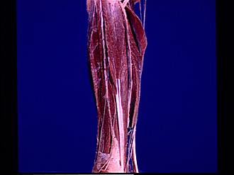
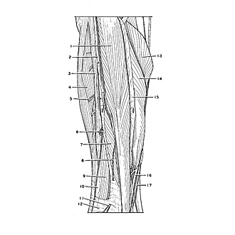
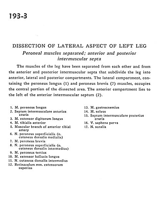
Dissection of lateral aspect of left leg
Peroneal muscles separated; anterior and posterior intermuscular septa
Image #193-3
KEYWORDS: Leg, Muscles and tendons.
Creative Commons
Stanford holds the copyright to the David L. Bassett anatomical images and has assigned Creative Commons license Attribution-Share Alike 4.0 International to all of the images.

For additional information regarding use and permissions, please contact the Medical History Center.
Dissection of lateral aspect of left leg
Peroneal muscles separated; anterior and posterior intermuscular septa
The muscles of the leg have beem separated from each other and from the anterior and posterior intermuscular septa that subdivide the leg into anterior, lateral and posterior compartments. The lateral compartment, containing the peroneus longus (1) and peroneus brevis (7) muscles, occupies the central portion of the dissected area. The anterior compartment lies to the left of the anterior intermuscular septum (2).
- Peroneus longus muscle
- Anterior intermuscular septum
- Extensor digitorum longus muscle
- Tibialis anterior muscle
- Muscular branch of anterior tibial artery
- Superficial peroneal nerve (dorsal intermediate cutaneous nerve)
- Peroneus brevis muscle
- Superficial peroneal nerve (dorsal medial cutaneous nerve)
- Peroneus tertius muscle
- Extensor hallucis longus muscle
- Dorsal intermediate cutaneous nerve
- Extensor retinaculum
- Gastrocnemius muscle
- Soleus muscle
- Posterior intermuscular septum
- Lesser saphenous vein
- Sural nerve