Bassett Collection of Stereoscopic Images of Human Anatomy
Dissection of anterior aspect of left leg
Anterior tibial and dorsal pedal vessels; anterolateral view of leg and foot
Image #192-7
KEYWORDS: Foot and toes, Leg, Muscles and tendons, Vasculature.
Creative Commons
Stanford holds the copyright to the David L. Bassett anatomical images and has assigned Creative Commons license Attribution-Share Alike 4.0 International to all of the images.

For additional information regarding use and permissions, please contact the Medical History Center.
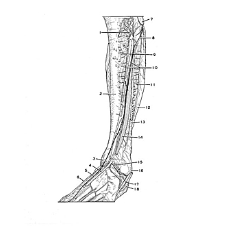
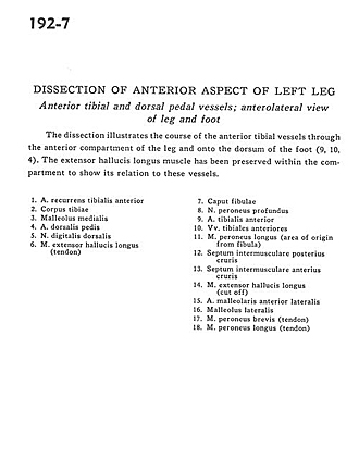
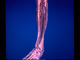
Dissection of anterior aspect of left leg
Anterior tibial and dorsal pedal vessels; anterolateral view of leg and foot
The dissection illustrates the course of the anterior tibial vessels through the anterior compartment of the leg and oto the dorsum of the foot (9,10,4). The extensor hallucis longus muscle has been preserved within the compartment to show its relation to these vessels.
- Anterior tibial recurrent artery
- Body of tibia
- Medial malleolus
- Dorsalis pedis artery
- Dorsal digital nerve
- Extensor hallucis longus muscle (tendon)
- Head of fibula
- Deep peroneal nerve
- Anterior tibial artery
- Anterior tibial veins
- Peroneus longus muscle (area of origin from fibula)
- Posterior intermuscular septum
- Anterior intermuscular septum
- Extensor hallucis longus muscle (cut off)
- Anterior lateral malleolar artery
- Lateral malleolus
- Peroneus brevis muscle (tendon)
- Peroneus longus muscle (tendon)