Bassett Collection of Stereoscopic Images of Human Anatomy
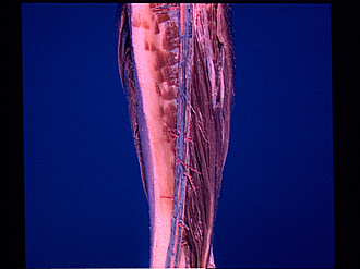
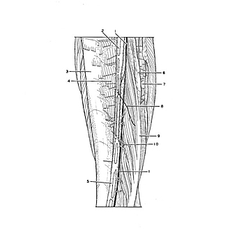
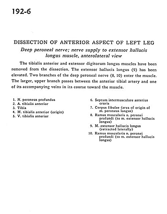
Dissection of anterior aspect of left leg
Deep peroneal nerve; nerve supply to extensor hallucis longus muscle, anterolateral view
Image #192-6
KEYWORDS: Leg, Muscles and tendons, Peripheral nervous system, Vasculature.
Creative Commons
Stanford holds the copyright to the David L. Bassett anatomical images and has assigned Creative Commons license Attribution-Share Alike 4.0 International to all of the images.

For additional information regarding use and permissions, please contact the Medical History Center.
Dissection of anterior aspect of left leg
Deep peroneal nerve; nerve supply to extensor hallucis longus muscle, anterolateral view
The tibialis anterior and extensor digitorum longus muscles have been removed from the dissection. The extensor hallucis longus (9) has been elevated. Two branches of the deep peroneal nerve (8, 10) enter the muscle. The larger, upper branch passes between the anterior tibial artery and one of its accompanying veins in its course toward the muscle.
- Deep peroneal nerve
- Anterior tibial artery
- Tibia
- Tibialis anterior muscle (origin)
- Anterior tibial vein
- Anterior intermuscular septum
- Body of fibula (area of origin of peroneus longus muscle)
- Muscular branch of deep peroneal nerve (to extensor hallucis longus muscle)
- Extensor hallucis longus muscle (retracted laterally)
- Muscular branch of deep peroneal nerve (to extensor hallucis longus muscle)