Bassett Collection of Stereoscopic Images of Human Anatomy
Dissection of anterior aspect of left leg
Nerve supply to extensor digitorum longus, close-up view of upper part of leg
Image #192-3
KEYWORDS: Leg, Muscles and tendons, Peripheral nervous system, Vasculature.
Creative Commons
Stanford holds the copyright to the David L. Bassett anatomical images and has assigned Creative Commons license Attribution-Share Alike 4.0 International to all of the images.

For additional information regarding use and permissions, please contact the Medical History Center.

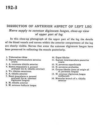
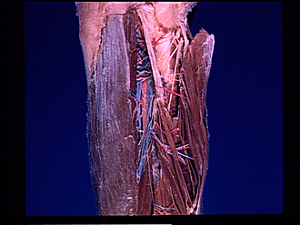
Dissection of anterior aspect of left leg
Nerve supply to extensor digitorum longus, close-up view of upper part of leg
This close-up photograph of the upper part of the leg the details of the blood vessels and nerves within the anterior compartment of the leg are clearly visible. Nerves that enter the extensor digitorum longus have been preserved in reflecting the muscle posteriorly.
- Tibial tuberosity
- Anterior intermuscular septum
- Anterior tibial recurrent artery
- Muscular branch of deep peroneal nerve (to tibialis anterior muscle)
- Anterior tibial veins
- Anterior tibial artery
- Muscular branch of deep peroneal nerve (to extensor digitorum longus muscle)
- Tibialis anterior muscle
- Extensor hallucis longus muscle
- Head of fibula
- Posterior intermuscular septum
- Superficial peroneal nerve
- Deep peroneal nerve
- Body of fibula (area of origin of peroneus longus muscle)
- Extensor digitorum longus muscle (reflected)
- Muscular branch of anterior tibial artery