
Bassett Collection of Stereoscopic Images of Human Anatomy
Dissection of knee
Interior of right knee joint, medial view
Image #191-4
KEYWORDS: Bones joints cartilage, Knee, Muscles and tendons.
Creative Commons
Stanford holds the copyright to the David L. Bassett anatomical images and has assigned Creative Commons license Attribution-Share Alike 4.0 International to all of the images.

For additional information regarding use and permissions, please contact the Medical History Center.
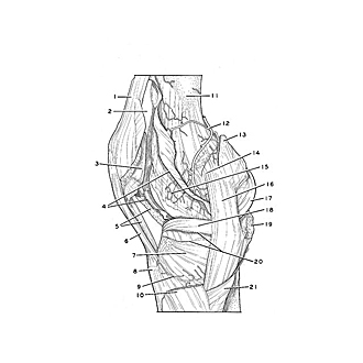
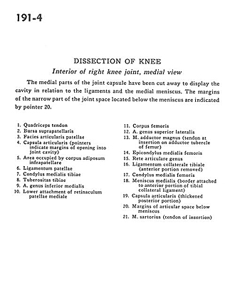
Dissection of knee
Interior of right knee joint, medial view
The medial parts of the joint capsule have been cut away to display the cavity in relation to the ligaments and the medial meniscus. The margins of the narrow part of the joint space located below the meniscus are indicated by pointer 20.
- Quadriceps tendon
- Suprapatellar bursa
- Patellar articular surface
- Articular capsule (pointers indicate margins of opening into joint cavity)
- Area occupied by infrapatellar fat body
- Patellar ligament
- Medial condyle of tibia
- Tibial tuberosity
- Medial inferior genicular artery
- Lower attachment of medial patellar retinaculum
- Body of femur
- Lateral superior genicular artery
- Adductor magnus muscle (tendon at insertion on adductor tubercle of femur)
- Medial epicondyle of femur
- Rete articulare genus
- Collateral ligament of tibia (anterior portion removed)
- Medial condyle of femur
- Medial meniscus (border attached to anterior portion of medial collateral ligament)
- Articular capsule (thickened posterior portion)
- Margins of articular space below meniscus
- Sartorius muscle (tendon of insertion)