Bassett Collection of Stereoscopic Images of Human Anatomy
Dissection of knee
Interior of right knee joint, cruciate ligaments divided
Image #190-6
KEYWORDS: Bones joints cartilage, Knee, Muscles and tendons.
Creative Commons
Stanford holds the copyright to the David L. Bassett anatomical images and has assigned Creative Commons license Attribution-Share Alike 4.0 International to all of the images.

For additional information regarding use and permissions, please contact the Medical History Center.

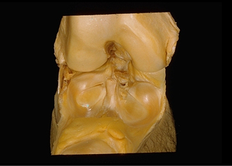
Dissection of knee
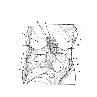
Interior of right knee joint, cruciate ligaments divided
The collateral ligaments and cruciate ligaments have been cut through to permit the separation of the femur from the tibia.
- Posterior cruciate ligament (divided)
- Anterior cruciate ligament (divided)
- Lateral condyle of femur
- Collateral ligament of fibula (divided)
- Iliotibial tract (cut off)
- Lateral meniscus (note its separation from fibular collateral ligament)
- Lateral condyle of tibia (articular surface)
- Infrapatellar fat body
- Patellar articular surface
- Line of reflection of articular capsule
- Medial condyle of femur
- Collateral ligament of tibia (divided)
- Upper pointer: Posterior meniscofemoral ligament (cut off) Lower pointer: Anterior meniscofemoral ligament (cut off)
- Medial meniscus (note fusion with tibial collateral ligament)
- Medial condyle of tibia (articular surface)