Bassett Collection of Stereoscopic Images of Human Anatomy
Dissection of knee
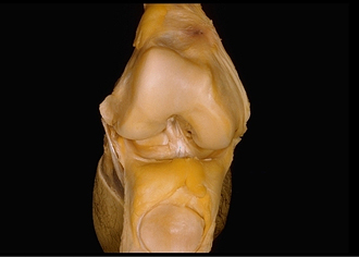
Interior of right knee joint, anterior cruciate ligament
Image #190-5
KEYWORDS: Bones joints cartilage, Knee, Muscles and tendons.
Creative Commons
Stanford holds the copyright to the David L. Bassett anatomical images and has assigned Creative Commons license Attribution-Share Alike 4.0 International to all of the images.

For additional information regarding use and permissions, please contact the Medical History Center.


Dissection of knee
Interior of right knee joint, anterior cruciate ligament
The infrapatellar synovial fold (12) has been cut off to reveal the anterior cruciate ligament (9). Synovial membrane partially encloses the cruciate ligament.
- Patellar femoral surface
- Lateral epicondyle
- Lateral condyle of femur
- Superior articular surface of tibia
- Lateral meniscus
- Infrapatellar fat body
- Patellar articular surface
- Suprapatellar bursa
- Anterior cruciate ligament
- Medial condyle of femur
- Medial meniscus
- Infrapatellar synovial fold (cut off)