Bassett Collection of Stereoscopic Images of Human Anatomy
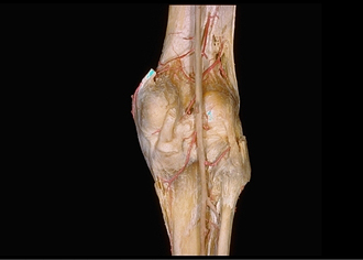
Dissection of knee
Capsule, ligaments and arteries of right knee, posterior view
Image #190-2
KEYWORDS: Knee, Muscles and tendons.
Creative Commons
Stanford holds the copyright to the David L. Bassett anatomical images and has assigned Creative Commons license Attribution-Share Alike 4.0 International to all of the images.

For additional information regarding use and permissions, please contact the Medical History Center.
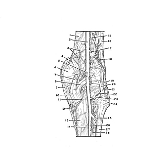

Dissection of knee
Capsule, ligaments and arteries of right knee, posterior view
- Body of femur
- Popliteal artery
- Lateral superior genicular arteries (duplicated)
- Adductor magnus muscle (tendon of insertion)
- Medial genicular artery (passing into intercondylar fossa)
- Medial condyle of femur
- Semimembranosus muscle (tendon of insertion)
- Oblique popliteal ligament
- Collateral ligament of tibia
- Medial condyle of tibia
- Lateral inferior genicular artery
- Semitendinosus muscle (tendon of insertion)
- Body of tibia
- Soleal line
- Muscular branch of popliteal artery
- Iliotibial tract
- Descending genicular artery (cut off)
- Medial superior genicular artery
- Collateral ligament of fibula
- Popliteal arcuate ligament
- Biceps fern oris muscle (tendon of insertion)
- Popliteus muscle (tendon of origin)
- Medial inferior genicular artery
- Head of fibula
- Anterior tibial artery
- Posterior tibial artery
- Body of fibula
- Interosseous membrane of leg