
Bassett Collection of Stereoscopic Images of Human Anatomy
Dissection of knee
Capsule, ligaments and arteries of right knee, anterior view
Image #189-7
KEYWORDS: Bones joints cartilage, Knee, Muscles and tendons.
Creative Commons
Stanford holds the copyright to the David L. Bassett anatomical images and has assigned Creative Commons license Attribution-Share Alike 4.0 International to all of the images.

For additional information regarding use and permissions, please contact the Medical History Center.
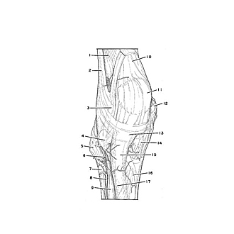
Dissection of knee
Capsule, ligaments and arteries of right knee, anterior view
The superficial structures and the muscles have been cut away to reveal the fibrous reinforcements of the anterior aspect of the right knee.
- Vastus lateralis muscle
- Iliotibial tract
- Lateral patellar retinaculum
- Lateral condyle of tibia
- Head of fibula
- Anterior tibial recurrent artery
- Body of fibula
- Anterior tibial artery
- Interosseous membrane of leg
- Quadriceps tendon
- Patella
- Medial epicondyle of femur
- Patellar ligament
- Medial condyle of tibia
- Tibial tuberosity
- Crural fascia
- Anterior border of tibia