
Bassett Collection of Stereoscopic Images of Human Anatomy
Dissection of anterior and medial aspects of thigh
Nerve supply to left vastus medialis, anteromedial view
Image #188-1
KEYWORDS: Muscles and tendons, Peripheral nervous system, Thigh.
Creative Commons
Stanford holds the copyright to the David L. Bassett anatomical images and has assigned Creative Commons license Attribution-Share Alike 4.0 International to all of the images.

For additional information regarding use and permissions, please contact the Medical History Center.
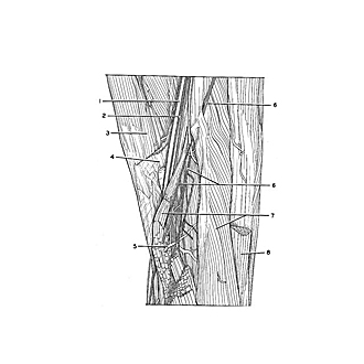
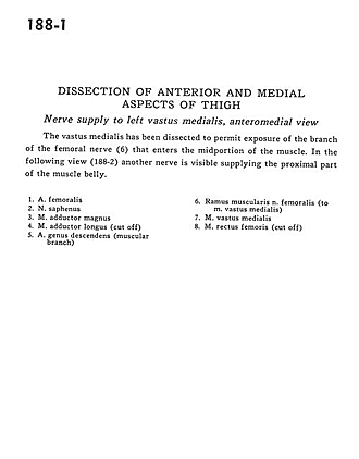
Dissection of anterior and medial aspects of thigh
Nerve supply to left vastus medialis, anteromedial view
The vastus medialis has been dissected to permit exposure of the branch of the femoral nerve (6) that enters the midportion of the muscle. In the following view (188-2) another nerve is visible supplying the proximal part of the muscle belly.
- Femoral artery
- Saphenous nerve
- Adductor magnus muscle
- Adductor longus muscle (cut off)
- Descending genicular artery (muscular branch)
- Muscular branch of femoral nerve (to vastus medialis muscle)
- Vastus medialis muscle
- Rectus femoris muscle (cut off)