Bassett Collection of Stereoscopic Images of Human Anatomy
Dissection of anterior and medial aspects of thigh
Deep femoral artery and perforating arteries of left thigh viewed from medial side
Image #187-7
KEYWORDS: Thigh, Vasculature.
Creative Commons
Stanford holds the copyright to the David L. Bassett anatomical images and has assigned Creative Commons license Attribution-Share Alike 4.0 International to all of the images.

For additional information regarding use and permissions, please contact the Medical History Center.
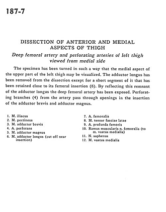
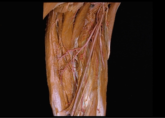
Dissection of anterior and medial aspects of thigh
Deep femoral artery and perforating arteries of left thigh viewed from medial side
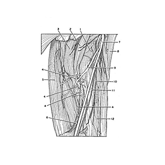
The specimen has been turned in such a way that the medial aspect of the upper part of the left thigh may be visualized. The adductor longus has been removed from the dissection except for a short segment of it has been removed from the dissection except for a short segment of it that has been retained close to its femoral insertion (6). By reflecting this remnant of the adductor longus the deep femoral artery has been exposed. Perforating branches (4) from the artery pass through the openings in the insertion of the adductor brevis and adductor magnus.
- Iliacus muscle
- Pectineus muscle
- Adductor brevis muscle
- Perforating artery
- Adductor magnus muscle
- Adductor longus muscle (cut off near insertion)
- Femoral artery
- Tensor fascia lata muscle
- Deep femoral artery
- Muscular branch of femoral nerve (to vastus medialis muscle)
- Saphenous nerve
- Vastus medialis muscle