Bassett Collection of Stereoscopic Images of Human Anatomy
Dissection of anterior and medial aspects of thigh
Nerve supply to lower part of sartorius; saphenous nerve; adductor canal and adductor hiatus, medial view
Image #187-4
KEYWORDS: Muscles and tendons, Peripheral nervous system, Thigh.
Creative Commons
Stanford holds the copyright to the David L. Bassett anatomical images and has assigned Creative Commons license Attribution-Share Alike 4.0 International to all of the images.

For additional information regarding use and permissions, please contact the Medical History Center.
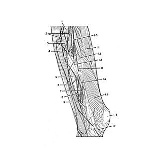
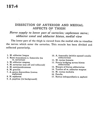
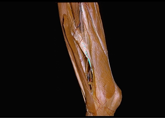
Dissection of anterior and medial aspects of thigh
Nerve supply to lower part of sartorius; saphenous nerve; adductor canal and adductor hiatus, medial view
The lower part of the thigh is viewed from the medial side to visualize the nerves which enter the sartorius. This muscle has been divided and reflected posteriorly.
- Adductor longus muscle
- Muscular branch femoral nerve (to sartorius muscle)
- Adductor magnus muscle
- Sartorius muscle (cut off and reflected)
- Semimembranosus muscle
- Gracilis muscle
- Descending genicular artery (saphenous branch)
- Saphenous nerve
- Popliteal artery (in background)
- Femoral artery (within opened adductor canal)
- Rectus femoris muscle
- Fascia bridging across hiatus adductorius
- Margin of hiatus adductorius
- Tendon of adductor magnus muscle
- Vastus medialis muscle
- Patella
- Infrapatellar branch of saphenous nerve