Bassett Collection of Stereoscopic Images of Human Anatomy
Dissection of anterior and medial aspects of thigh
Adductor longus and pectineus muscles
Image #187-1
KEYWORDS: Muscles and tendons, Thigh.
Creative Commons
Stanford holds the copyright to the David L. Bassett anatomical images and has assigned Creative Commons license Attribution-Share Alike 4.0 International to all of the images.

For additional information regarding use and permissions, please contact the Medical History Center.
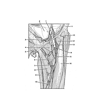
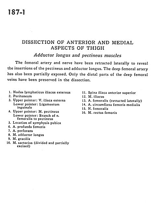
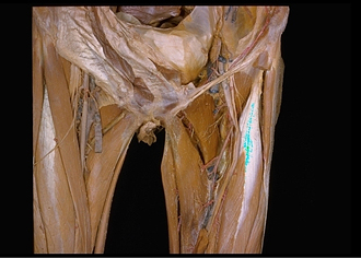
Dissection of anterior and medial aspects of thigh
Adductor longus and pectineus muscles
The femoral artery and nerve have been retracted laterally to reveal the insertions of the pectineus and adductor longus. The deep femoral artery has also been partially exposed. Only the distal parts of the deep femoral veins have been preserved in the dissection.
- External iliac lymph node
- Peritoneum
- Upper pointer: External iliac vein Lower pointer: Inguinal ligament
- Upper pointer: Pectineus muscle Lower pointer: Branch of femoral nerve to pectineus
- Location of pubic symphysis
- Deep femoral artery
- Perforating artery
- Adductor longus muscle
- Gracilis muscle
- Sartorius muscle (divided and partially excised)
- Anterior superior iliac spine
- Iliacus muscle
- Femoral artery (retracted laterally)
- Medial femoral circumflex artery
- Femoral nerve
- Rectus femoris muscle