
Bassett Collection of Stereoscopic Images of Human Anatomy
Dissection of anterior and medial aspects of thigh
Anterior branches of obturator nerve and vessels in relation to adductor longus
Image #186-6
KEYWORDS: Muscles and tendons, Peripheral nervous system, Thigh, Vasculature.
Creative Commons
Stanford holds the copyright to the David L. Bassett anatomical images and has assigned Creative Commons license Attribution-Share Alike 4.0 International to all of the images.

For additional information regarding use and permissions, please contact the Medical History Center.

Dissection of anterior and medial aspects of thigh
Anterior branches of obturator nerve and vessels in relation to adductor longus
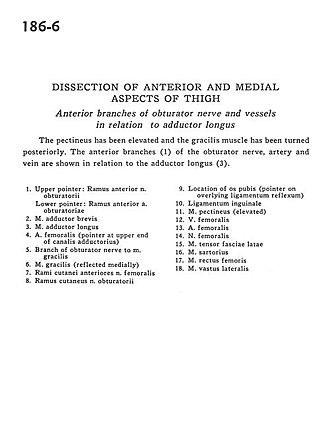
The pectineus has been elevated and the gracilis muscle has been turned posteriorly. The anterior branches (1) of the obturator nerve, artery and vein are shown in relation to the adductor longus (3).
- Upper pointer: Anterior branch of obturator nerve Lower pointer: Anterior branch of obturator artery
- Adductor brevis muscle
- Adductor longus muscle
- Femoral artery (pointer at upper end of adductor canal)
- Branch of obturator nerve to gracilis muscle
- Gracilis muscle (reflected medially)
- Cutaneous branches of anterior femoral nerve
- Cutaneous branch of obturator nerve
- Location of pubic bone (pointer on overlying reflex inguinal ligament)
- Inguinal ligament
- Pectineus muscle (elevated)
- Femoral vein
- Femoral artery
- Femoral nerve
- Tensor fasciae latae muscle
- Sartorius muscle
- Rectus femoris muscle
- Vastus lateralis muscle