
Bassett Collection of Stereoscopic Images of Human Anatomy
Dissection of anterior and medial aspects of thigh
Femoral sheath
Image #186-2
KEYWORDS: Fascia, Muscles and tendons, Thigh.
Creative Commons
Stanford holds the copyright to the David L. Bassett anatomical images and has assigned Creative Commons license Attribution-Share Alike 4.0 International to all of the images.

For additional information regarding use and permissions, please contact the Medical History Center.
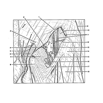

Dissection of anterior and medial aspects of thigh
Femoral sheath
Below the inguinal ligament the femoral sheath (13) has been exposed by removing the superficial inguinal lymph nodes, the cribiform fascia and part of the fascia lata which bounds the saphenous opening. The inferior part of the falciform margin of this opening has been retained (17).
- Inguinal ligament
- Location of femoral canal (note lymphatic vessels penetrating femoral sheath in this area) 3. Intercrural fibers
- Superficial inguinal ring (left pointer, medial crus; right pointer, lateral crus)
- Ilioinguinal nerve (cut off)
- Spermatic cord (external spermatic fascia removed cremasteric fascia exposed)
- Pectineus muscle (covered by fascia)
- Adductor longus muscle (covered by fascia lata)
- Lateral femoral cutaneous nerve
- Iliopsoas muscle (covered by iliopsoas fascia)
- Cutaneous branches of anterior femoral nerve
- Superficial circumflex iliac artery
- Femoral sheath
- Sartorius muscle (covered by fascia lata)
- Greater saphenous vein (cut off)
- Deep inguinal lymph node
- Margo falciformis (inferior horn)
- Fascia lata