Bassett Collection of Stereoscopic Images of Human Anatomy
Dissection of anterior and medial aspects of thigh
General view of superficial vessels and nerves of left thigh; anterior muscles of right thigh
Image #185-3
KEYWORDS: Muscles and tendons, Peripheral nervous system, Thigh, Vasculature.
Creative Commons
Stanford holds the copyright to the David L. Bassett anatomical images and has assigned Creative Commons license Attribution-Share Alike 4.0 International to all of the images.

For additional information regarding use and permissions, please contact the Medical History Center.

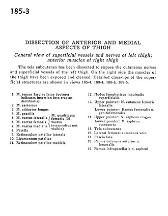
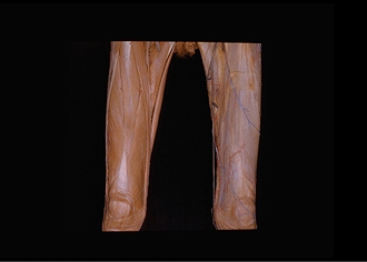
Dissection of anterior and medial aspects of thigh
General view of superficial vessels and nerves of left thigh; anterior muscles of right thigh
The tela subcutanea has been dissected to expose the cutaneous nerves and superficial vessels of the left thigh. On the right side the muscles of the thigh have been exposed and cleaned. Detailed close-ups of the superficial structures are shown in views 185-4, 189-4, 189-5, 189-6.
- Tensor fasciae latae muscle (pointer indicates insertion into Iliotibial tract)
- Sartorius muscle
- Adductor longus muscle
- Gracilis muscle
- Vastus lateralis muscle
- Rectus femoris muscle
- Vastus medialis muscle
- Patella
- Lateral patellar retinaculum
- Patellar ligament
- Medial patellar retinaculum
- Superficial inguinal lymph node
- Upper pointer: Lateral femoral cutaneous nerve Lower pointer: Femoral branch of genitofemoral nerve
- Upper pointer: Greater saphenous vein Lower pointer: Accessory saphenous vein
- Tela subcutanea
- Lateral femoral cutaneous vein
- Fascia lata
- Anterior cutaneous branch of femoral nerve
- Infrapatellar branch of saphenous nerve