Bassett Collection of Stereoscopic Images of Human Anatomy
Dissection of iliopsoas muscle and lumbar plexus
Lumbar plexus; nerves to psoas major
Image #184-7
KEYWORDS: Muscles and tendons, Peripheral nervous system.
Creative Commons
Stanford holds the copyright to the David L. Bassett anatomical images and has assigned Creative Commons license Attribution-Share Alike 4.0 International to all of the images.

For additional information regarding use and permissions, please contact the Medical History Center.
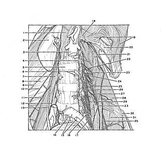
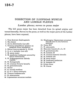
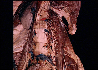
Dissection of iliopsoas muscle and lumbar plexus
Lumbar plexus; nerves to psoas major
The left psoas major has been detached from its spinal origins and turned laterally. Nerves to the psoas, as well as the major parts of the lumbar plexus, have been exposed.
- Right crus of diaphragm
- Right kidney
- Abdominal aorta (cut off)
- Sympathetic trunk (pointer on sympathetic trunk ganglia)
- Intervertebral disc L. II-III
- Lumbar artery and vein (cut off)
- Psoas major muscle
- Quadratus lumborum muscle covered by transversalis fascia
- Iliohypogastric nerve (faintly visible)
- Ilioinguinal nerve
- Lateral femoral cutaneous nerve
- Genitofemoral nerve
- Ureter
- Middle sacral artery
- Common iliac artery and vein
- Lumbosacral trunk
- Obturator nerve
- Medial arcuate ligament
- Diaphragm (Lateral arcuate ligament lies in area between pointers)
- Rib XII
- Subcostal nerve
- Muscular branch of nerve L. II
- Genitofemoral nerve
- Lumbar nerve II
- Lateral femoral cutaneous nerve
- Lumbar nerve III
- Psoas major muscle
- Quadratus lumborum muscle
- Lumbar nerve IV
- Iliac crest
- Iliacus muscle