Bassett Collection of Stereoscopic Images of Human Anatomy
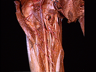
Dissection of posterior aspect of left thigh
Nerve supply to hamstring muscles
Image #183-7
KEYWORDS: Muscles and tendons, Peripheral nervous system, Thigh, Vasculature.
Creative Commons
Stanford holds the copyright to the David L. Bassett anatomical images and has assigned Creative Commons license Attribution-Share Alike 4.0 International to all of the images.

For additional information regarding use and permissions, please contact the Medical History Center.
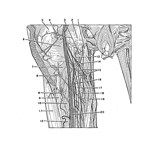

Dissection of posterior aspect of left thigh
Nerve supply to hamstring muscles
The biceps and semitendinosus muscles have been detached from their origins on the ischial tuberosity to permit them to be dissected. Branches of the tibial nerve and of the medial circumflex femoral artery enter the proximal parts of the muscle bellies.
- Tendons of origin of biceps femoris and semitendinosus (detached)
- Ischial tuberosity
- Adductor magnus muscle (origin)
- Ascending branch of medial femoral circumflex artery
- Greater trochanter
- Quadratus femoris muscle
- Adductor minimus muscle
- Perforating artery
- Lateral intermuscular septum
- Adductor magnus muscle (insertion)
- Vastus lateralis muscle
- Short head of biceps femoris muscle
- Medial femoral circumflex artery (deep branch)
- Muscular branch of sciatic nerve (to semitendinosus muscle)
- Muscular branch of sciatic nerve (to long head of biceps femoris)
- Muscular branch of sciatic nerve (to semimembranosus muscle)
- Semimembranosus muscle
- Semitendinosus muscle
- Long head of biceps femoris
- Sciatic nerve